Your Cart is Empty
Start shopping
IGF-1 DES vs IGF-1 LR3: Truncated vs Extended Variants in Anabolic Signaling Research
David Fuller
Last Updated On:October 8, 2026
Compare IGF-1 DES vs IGF-1 LR3 for anabolic signaling research: IGFBP binding, half-life around 20–30 hours, and choosing local vs systemic variants for in vitro or in vivo work.

IGF-1 DES and IGF-1 LR3 are engineered analogs of insulin-like growth factor 1 that have become indispensable tools in preclinical muscle physiology and anabolic signaling research. IGF-1 DES achieves its effects through N-terminal truncation, the deletion of three amino acids that normally interact with binding proteins. The result is a hyperlocal, highly potent peptide well suited to cell-autonomous hypertrophy work.
On the other hand, IGF-1 LR3 gains extended systemic activity through arginine substitution and an N-terminal extension that prolongs half-life and enables whole-organism investigation of anabolic signaling. Both peptides activate the same IGF-1 receptor and drive the same downstream growth pathways, yet their distinct pharmacokinetic and bioavailability profiles create fundamentally different research contexts.
This IGF-1 DES vs LR3 comparison walks through the mechanistic divergence, the practical protocol considerations, and the research applications that separate them, so you can select the variant that matches your experimental question.
IGF-1 Variants in Anabolic Signaling Research
The insulin-like growth factor 1 (IGF-1) family is one of the most extensively studied pathways in muscle growth & performance peptides research and cellular proliferation work. Within this category, two synthetic variants have emerged as the primary tools for preclinical investigation: IGF-1 DES and IGF-1 LR3. Their mechanistic differences shape distinct research applications, and the choice between them hinges not on which is “superior” but on whether your endpoint requires intense local action or sustained systemic presence.
Both peptides activate the IGF-1 receptor, but they arrive at that activation through different biochemical routes. Different handling by binding proteins, circulation half-lives, and tissue-penetration profiles mean a protocol optimized for one variant may yield inadequate or misleading results with the other. For rigorous comparative design, the mechanistic detail matters.
IGF-1 DES: Truncation, IGFBP Binding, and Local Potency
IGF-1 DES (des[1-3]-IGF-1) is a truncated variant lacking the first three N-terminal amino acids present in wild-type IGF-1 [4]. That simple deletion has outsized biochemical consequences.
The primary effect is ablation of binding to insulin-like growth factor-binding proteins (IGFBPs). Wild-type IGF-1 circulates primarily in ternary complexes with IGFBP-3 (and to a lesser extent IGFBP-5) plus acid-labile subunit (ALS) [1]. This configuration protects the peptide from degradation but limits tissue bioavailability. When IGF-1 DES loses those three amino acids, its affinity for IGFBPs falls sharply.
Research on IGFBP structure and IGF-binding interactions shows that des-(1–3)-IGF-I binds to IGFBP-3 with several-fold lower affinity than natural IGF-I, with similarly reduced binding to other IGFBPs [1]. Relative to native IGF-1, the peptide becomes largely unbound and bioavailable, a property that confers both advantages and experimental constraints.
This truncation creates a locally potent, free-acting variant. In cultured myotubes and satellite cells, IGF-1 DES exhibits sharp dose-response curves and rapid activation of downstream pathways. Without IGFBP sequestration, higher local concentrations accumulate quickly at the site of administration, whether that is a perfused cell culture dish or an injected rodent muscle. The peptide does not compete for IGFBP binding slots and does not rely on circulating carrier proteins for transport. What you add is what acts.
That profile makes IGF-1 DES a preferred choice for in vitro work where local potency and clean dose-response relationships are priorities. The unbound nature simplifies interpretation: changes in myogenic differentiation or proliferation correlate directly to the applied concentration, without the confounding variable of variable binding protein availability.
In vivo, the freedom comes with a caveat. The circulating half-life, measured in minutes, means systemic IGF-1 DES concentrations drop rapidly after administration. Maintaining anabolic stimulation in intact organisms requires frequent dosing or sustained local delivery, which is why preclinical muscle hypertrophy studies using IGF-1 DES often employ direct intramuscular injection. That route leverages the local concentration advantage but sidesteps the challenge of maintaining systemic levels.
IGF-1 LR3: Arginine Substitution, Extended Half-Life, and Systemic Action
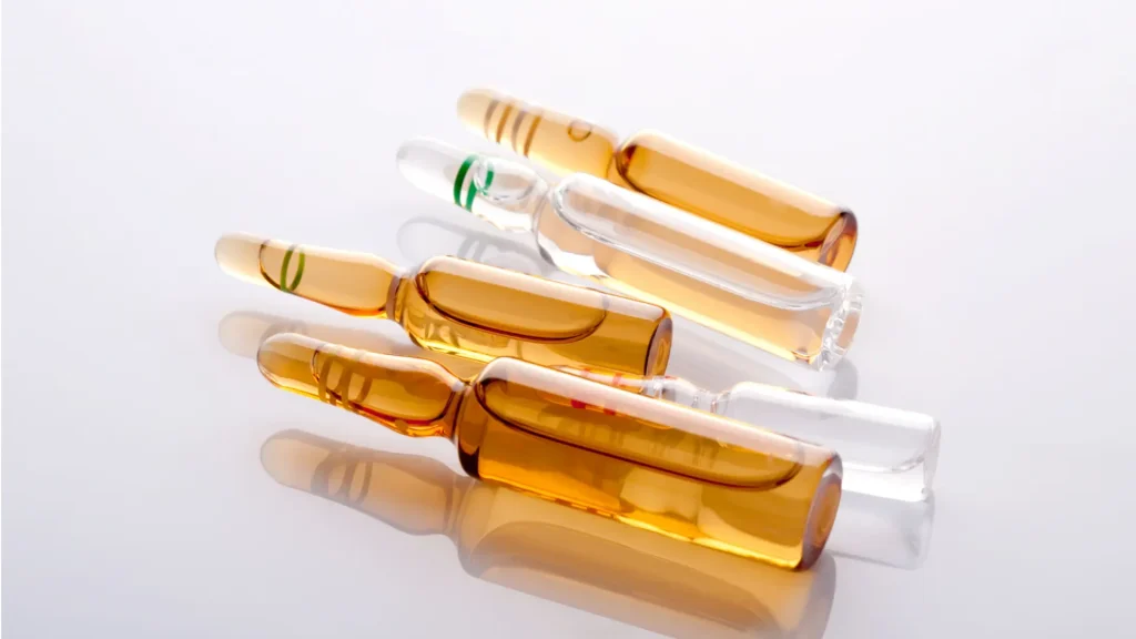
IGF-1 LR3 (long-R3-IGF-1) takes a different approach. Rather than deletion, it substitutes the third amino acid (glutamic acid to arginine) and adds a 13-residue N-terminal extension [6]. Together, these modifications confer a critical advantage: heavy resistance to IGFBP binding, with retained circulation.
The arginine substitution reduces affinity for IGFBPs by over 1,000-fold relative to native IGF-1, making IGF-1 LR3 largely resistant to IGFBP sequestration [5]. As a result, it has markedly greater tissue bioavailability than the native peptide while maintaining systemic circulation.
The dominant pharmacokinetic feature of IGF-1 LR3 is its extended half-life. In rodent models, IGF-1 LR3 persists in circulation for many hours, with an elimination half-life commonly reported around 20–30 hours [5]. This is far longer than native IGF-1 or IGF-1 DES. A single injection of IGF-1 LR3 can sustain receptor activation across target tissues for a prolonged period, making it the natural choice for whole-animal studies that examine tissue response to continuous or semi-continuous IGF-1 receptor stimulation without repeated dosing.
The extended half-life also carries a research design advantage. It dampens the noise of minute-to-minute variation in local peptide concentration. In IGF-1 DES protocols, especially those relying on bolus dosing, transient peaks and troughs can complicate kinetic analysis. IGF-1 LR3’s pharmacokinetics favor smoother exposure profiles and more stable downstream signaling over time.
Receptor Binding Affinity and Downstream PI3K/Akt/mTOR Activation
Both IGF-1 DES and IGF-1 LR3 bind the type 1 IGF receptor (IGF-1R) with high affinity and activate the classical anabolic cascade: receptor autophosphorylation, PI3K recruitment, Akt phosphorylation, and downstream mTORC1 activation via TSC1/2 inhibition [7]. Foundational research on this pathway demonstrates that Akt/mTOR signaling is a central regulator of skeletal muscle hypertrophy in vivo [3].
At the receptor level, the two variants show comparable potency, with maximal IGF-1R activation reached at nanomolar concentrations. Affinity constants differ slightly, and IGF-1 LR3 may show marginally reduced binding relative to wild-type or DES in some assay systems. The practical significance is minimal, since both reach saturation at equivalent physiological concentrations.
They diverge in the temporal and spatial profile of activation. IGF-1 DES, with its rapid local accumulation and high local concentration, generates sharp, steep dose-response curves in cell-based assays. IGF-1 LR3’s extended half-life and more gradual tissue penetration produce gentler activation curves but sustained pathway engagement.
Downstream, both peptides engage Akt through PI3K-dependent pathways, leading to mTORC1 phosphorylation of S6K and 4E-BP1, the canonical drivers of protein translation initiation [7]. Both also suppress GSK3β and FoxO transcription factors via Akt, reducing protein degradation. The endpoint is consistent: net protein synthesis favorable to an anabolic state.
The practical distinction emerges in study design. If your research question centers on the dose-response relationship of a specific effector to IGF-1 stimulation, IGF-1 DES’s linear, concentration-dependent activation may provide cleaner data. If the question concerns sustained pathway engagement and tissue-level phenotypes over days to weeks in vivo, IGF-1 LR3’s pharmacokinetic profile is the better match.
In Vitro vs In Vivo Considerations for Each Variant
In vitro, IGF-1 DES dominates. Culture systems lack the IGFBP pool present in serum or plasma, so the truncated variant’s freedom from IGFBP binding is an asset rather than a liability. Dose-response relationships are clean, and pathway activation is attributable directly to peptide concentration. Cell lines and primary myotubes treated with IGF-1 DES show robust, reproducible hypertrophy and proliferation across a broad range of experimental protocols.
The absence of IGFBP binding also means IGF-1 DES is less susceptible to IGFBP-mediated feedback, a known confound in longer-running culture. Some in vitro systems show that IGFBP accumulation, especially from the cells themselves, can blunt IGF-1 signaling over time. IGF-1 DES sidesteps this issue entirely.
In vivo, the calculus shifts. Living tissue operates in the context of circulating IGFBPs, competing growth factors, hormonal milieu, and regional blood flow. IGF-1 DES’s short half-life demands either intramuscular injection to bypass systemic circulation or very frequent dosing. Neither is impractical for rodent models, but both narrow the experimental scope. By necessity, you are studying local muscle response to a high local IGF-1 DES concentration, not systemic IGF-1 action broadly.
Alternatively, IGF-1 LR3 circulates efficiently and can be administered by intraperitoneal or subcutaneous injection, reaching muscle, liver, adipose, and bone to elevate IGF-1 signaling across the whole animal [5]. This is the appropriate model for questions about systemic anabolic effects. Does elevated IGF-1 signaling improve recovery from disuse atrophy? How does chronic IGF-1 elevation affect metabolic rate? What are the off-target effects of sustained IGF-1R activation across tissues?
This makes the choice contextual:
- If you are investigating a cell-autonomous effect of IGF-1 on myogenic differentiation, IGF-1 DES in culture.
- If you are modeling systemic effects of elevated IGF-1 in an intact organism, IGF-1 LR3 in vivo.
Muscle Hypertrophy and Cell Proliferation Research Data

Both IGF-1 variants produce robust hypertrophy and proliferation in preclinical models, but the magnitude and kinetics differ in ways that align with their mechanistic profiles.
In myotubes, IGF-1 DES application produces rapid increases in myogenic protein content, often visible within 24 hours at physiological nanomolar doses. The magnitude of response scales linearly with concentration, which suits dose-escalation study designs. Recent work demonstrates how IGF-1 accelerates satellite cell differentiation and promotes myotube hypertrophy, with myogenic markers such as myosin heavy chain (MyHC) showing significant upregulation [2].
Published studies consistently show IGF-1 DES-induced hypertrophy accompanied by increased expression of hypertrophy-associated genes (myosin heavy chain, developmental myosin isoforms) and reduced expression of atrophy markers (MuRF1, MAFbx). In rodent in vivo studies, direct intramuscular IGF-1 DES injection produces localized muscle growth at the injection site. The effect is rapid and substantial, typically measurable within days, but it remains regionally confined. That regional constraint is mechanistically coherent, since the peptide does not circulate effectively and only muscle adjacent to the injection site receives high concentrations.
IGF-1 LR3 in vivo studies show more widespread effects across the musculature. Peer-reviewed experimental evidence confirms that IGF-1R signaling drives whole-animal anabolic responses to elevated IGF-1 in systemic investigation [3]. Systemic IGF-1 LR3 elevation via intraperitoneal injection increases whole-body lean mass and muscle cross-sectional area across multiple muscle groups. The effect typically needs slightly longer timescales, on the order of 1–2 weeks to reach significant magnitude, consistent with its pharmacokinetic profile.
For research aligned with body composition & recovery peptides applications, this longer timescale is an advantage. You are observing the response to systemic anabolic stimulation and sustained recovery rather than the response to supraphysiological local concentration.
One nuance worth flagging: in vivo IGF-1 LR3 studies often incorporate metabolic readouts such as body composition, glucose handling, and systemic insulin sensitivity, precisely because the variant reaches the liver, adipose, and other tissues beyond skeletal muscle. IGF-1 DES protocols, constrained to intramuscular injection, rarely report systemic metabolic effects. The question does not arise. That difference is not a failing of either design; it reflects the appropriate research applications of each peptide.
Protocol Considerations: Dosing Windows and Tissue Targeting
For IGF-1 DES in vitro work, standard practice ranges from 10 nM to 1 μM in acute dose-response experiments, with sustained stimulation at 100–500 nM in longer differentiation assays. Because the peptide is unbound, these are genuine free concentrations acting on the receptor. Incubation windows are short because IGF-1 DES is rapidly cleared in culture via receptor-mediated endocytosis and proteolysis [5]. Apparent stability depends on cell density, media composition, and incubation time. For longer storage, aliquot and cryopreserve peptides.
In vivo IGF-1 DES protocols typically employ intramuscular injection at doses ranging from 1 to 100 μg per injection, depending on rodent muscle mass and endpoint. Repeated dosing every 2–3 days is common to maintain anabolic stimulus, reflecting the short circulating half-life. Local muscle elevation at the injection site is dramatic, but that is the intent and the advantage for dissecting local myogenic responses.
IGF-1 LR3 in vivo dosing looks considerably different. Systemic IGF-1 LR3 protocols in rodents typically employ intraperitoneal injection at 100–300 μg/kg body weight, often dosed once or twice weekly. The extended half-life means less frequent dosing is required. The goal is steady-state elevation of circulating IGF-1 LR3 across the treatment window rather than transient local peaks.
For both variants, dose selection hinges on the study endpoint. Hypertrophy studies often use lower doses to model physiological elevation, while recovery-from-atrophy or regeneration studies may employ higher doses to accelerate kinetics. The key is justifying the dose against published preclinical literature and your specific research question.
Local vs Systemic: Choosing the Right IGF-1 Variant
IGF-1 DES fits investigations of cell-autonomous myogenic processes (differentiation, proliferation, hypertrophy at the myotube or myoblast level), concentration-response work where precise control of local peptide is essential, intracellular signaling mechanisms downstream of IGF-1R activation, local muscle response without systemic confounds, and protocols where rapid, sharp activation is preferable to sustained action.
IGF-1 LR3 suits whole-animal anabolic questions: responses to elevated IGF-1 signaling across multiple tissues (muscle, liver, adipose, bone), long-term recovery from atrophy, disuse or aging models, and metabolic integration of IGF-1 signaling with other hormones and nutrient status. This is also the context where researchers often study interactions with growth hormone peptides and other anabolic pathways. It fits protocols where sustained exposure over days to weeks reflects the biological question being asked.
In practice, a comprehensive program might employ both. Preliminary mechanistic work can use IGF-1 DES in cell culture to dissect the pathways and identify candidate effectors, and subsequent in vivo validation can use IGF-1 LR3 to confirm that the mechanism translates to systemic anabolic response. Many published studies of this design already exist. The two variants are complementary rather than competitors.
Mechanistic rigor, appropriate tool selection, and transparent sourcing are the foundations of reproducible IGF-1 research.
The content on this page is for informational and educational purposes only and is not intended as medical advice. Med Supply Solutions does not sell or distribute research peptides. All research must be conducted by qualified professionals in accordance with applicable laws and regulations.
Citations
[1] Baxter, R C. “Insulin-like growth factor (IGF)-binding proteins: interactions with IGFs and intrinsic bioactivities.” American journal of physiology. Endocrinology and metabolism vol. 278,6 (2000): E967-76. doi:10.1152/ajpendo.2000.278.6.E967
[2] Li, Xin et al. “Effect of IGF1 on Myogenic Proliferation and Differentiation of Bovine Skeletal Muscle Satellite Cells Through PI3K/AKT Signaling Pathway.” Genes vol. 15,12 1494. 21 Nov. 2024, doi:10.3390/genes15121494
[3] Yin, Lijun et al. “Crucial role of androgen receptor in resistance and endurance trainings-induced muscle hypertrophy through IGF-1/IGF-1R- PI3K/Akt- mTOR pathway.” Nutrition & metabolism vol. 17 26. 30 Mar. 2020, doi:10.1186/s12986-020-00446-y
[4] “Des(1-3)IGF-1.” Wikipedia, The Free Encyclopedia, Wikimedia Foundation, last edited 24 May 2026, en.wikipedia.org/wiki/Des(1-3)IGF-1.
[5] HealthRX.com Editorial Team. “IGF-1 LR3: Mechanisms, Dosing, Safety, and Clinical Evidence.” HealthRX.com, 15 Jan. 2025, healthrx.com/peptides-specialty/igf-1-lr3.
[6] “IGF-1 LR3.” Wikipedia, The Free Encyclopedia, Wikimedia Foundation, last edited 19 June 2023, en.wikipedia.org/wiki/IGF-1_LR3.
[7] Philippou, Anastassios, and Elisabeth R Barton. “Optimizing IGF-I for skeletal muscle therapeutics.” Growth hormone & IGF research : official journal of the Growth Hormone Research Society and the International IGF Research Society vol. 24,5 (2014): 157-63. doi:10.1016/j.ghir.2014.06.003